


 Tropilaelaps Mites
By: Clarence Collison
A potential future threat of the western honey bee (Apis mellifera) in North America are the parasitic mites in the genus Tropilaelaps which are currently confined to Asia and bordering areas. These mites are native brood parasites of the non-domesticated giant Asian honey bees, Apis dorsata, A. breviligula and A. laboriosa. They have spread onto the managed European honey bee (A. mellifera) sometime after humans introduced that bee into Asia. Nowadays, A. mellifera is kept for beekeeping throughout Asia and Tropilaelaps mites are one of its most damaging pests (Anderson and Roberts, 2013).
Mites of the genus Tropilaelaps (Acari: Laelapidae) are ectoparasites of honey bees native to Asia (Delfinado and Baker, 1961; Laigo and Morse, 1968). The primary host of one of the better known species Tropilaelaps clareae is Apis dorsata (Laigo and Morse, 1968) but Tropilaelaps mites were able to switch to the western honey bee, Apis mellifera (Delfinado and Baker, 1961; Anderson and Morgan, 2007). Tropilaelaps clareae was first discovered on A. mellifera in the Philippines (Delfinado and Baker, 1961). Tropilaelaps species seem to be prevalent in Asia and are able to infect a wide spectrum of honey bee species ranging from Apis mellifera, A. cerana, A. dorsata, A. florae and A. laboriosa (Bailey and Ball, 1991). However, these mites appear to be particularly pathogenic in A. mellifera (Burgett et al., 1983; De Jong et al., 1982; Laigo and Morse, 1969). Similar to Varroa destructor (Acari: Varroaidae), Tropilaelaps mites are infecting brood and suck hemolymph/fat from the body. Up to four female mites can invade the same brood cell (Burgett and Akratanakul, 1985; De Jong et al., 1982; Dainat et al., 2009).
Varroa and Tropilaelaps mites have coexisted in Apis mellifera (western honey bee) colonies in Asia for >50 years (Delfinado, 1963). However, Tropilaelaps mites are considered to be the more dominating and reproductively successful parasites of A. mellifera than Varroa mites (Burgett et al., 1983; Buawangpong et al., 2015). In 2007, a molecular examination of Tropilaelaps mites collected from different honey bee hosts from several Asian countries revealed two new Tropilaelaps species (Tropilaelaps mercedesae and Tropilaelaps thaii) distinctly separate from Tropilaelaps clareae and Tropilaelaps koenigerum (Anderson and Morgan, 2007). Among these four species, T. mercedesae and T. clareae are the most serious Tropilaelaps mites of A. mellifera. However, T. mercedesae exhibits a wider distribution than T. clareae. The life history of Tropilaelaps mites and food requirements are similar to that of Varroa mites. As a result, both mite genera can inflict severe damages on A. mellifera colonies (De Guzman et al., 2017).
An infestation by Tropilaelaps can be recognized either visually on bees and brood or by examining hive debris. Feeding on bee larvae and pupae causes brood malformation, death of bees and subsequent colony decline or absconding. Development requires about one week, and the mites are dispersed on bees. Irregular brood pattern, dead or malformed immatures, bees with malformed wings that crawl at the hive’s entrance and especially the presence of fast-running, large, red-brown, elongated mites on the combs, are diagnostic for the presence of T. clareae. An early diagnosis can be made by opening brood cells and finding immature and adult mites therein (Sammataro, 2004).
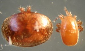
Varroa (left) & Tropilaelaps (right)
Combs of sealed honey bee worker brood (Apis mellifera ligustica hybrid) were taken from colonies infested by Tropilaelaps clareae and the numbers of mites in different developmental stages recorded. The duration of successive stages was estimated from the times at which the first and last individuals in each stage appeared and also from the relative frequency of occurrence of the different stages in the mite population of infested cells. Altogether 22 eggs, 42 larvae, 187 protonymphs, 251 deutonymphs and 659 young or old imagos (final and fully developed adult stage) were detected. The first individuals of successive stages in the development of the mite were found on honey bee brood of the following stages: mite eggs and larvae on spinning larvae, protonymphs on prepupae, deutonymphs on day-old pupae and young imagos on pupae four days old. The most advanced age of a developing bee (calculated from the time of egg-laying) with which each stage of mite was associated was: egg, 14 days; larvae, 15 days; protonymphs, 16 days and deutonymphs, 19 days. The calculated lengths of successive T. clareae stages were: eggs, 0.3–0.4 days; larvae, 0.3–0.6 days; protonymphs, 1.7–2.0 days and deutonymphs, 3.0-3.8 days. Length of the total developmental period was six days. All mites completed their development before the honey bees emerged, a factor that probably contributes to the faster population build-up of T. clareae than of Varroa jacobsoni (Woyke, 1987).
Tropilaelaps mercedesae parasitism can cause Apis mellifera colony mortality in Asia. Phokasem et al. (2019) reported that Tropilaelaps mites feed on both pre- and post-capped stages of honey bees. Feeding on pre-capped brood may extend their survival outside capped brood cells, especially in areas where brood production is year-round. They examined the types of injury inflicted by Tropilaelaps mites on different stages of honey bees, the survival of adult honey bees and level of honey bee viruses in fourth instar larvae and prepupae. The injuries inflicted on different developing honey bee stages were visualized by staining with trypan blue. Among pre-capped stages, fourth instar larvae sustained the highest number of wounds (4.6±0.5/larva) while second and third larval instars had at least two wounds. Consequently, wounds were evident on un-infested capped brood (fifth and sixth instar larvae=3.91±0.64 wounds; prepupae=5.25±0.73 wounds). Tropilaelaps mite infestations resulted in 3.4- and six-fold increases in the number of wounds in fifth and sixth instar larvae and prepupae as compared to un-infested capped brood, respectively. When wound inflicted prepupae metamorphosed to white-eyed pupae, all wound scars disappeared with the exuviae. This healing of wounds contributed to the reduction of the number of wounds (≤10) observed on the different pupal stages. Transmission of mite-borne virus such as Deformed Wing Virus (DWV) was also enhanced by mites feeding on early larval stages. DWV and Black Queen Cell Virus (BQCV) were detected in all fourth instar larvae and prepupae analyzed. However, viral levels were more pronounced in scarred fourth instar larvae and infested prepupae. The remarkably high numbers of wounds and viral load on scarred or infested developing honey bees may have caused significant weight loss and extensive injuries observed on the abdomen, wings, legs, proboscis and antennae of adult honey bees. Together, the survival of infested honey bees was significantly compromised. This study demonstrates the ability of Tropilaelaps mites to inflict profound damage on A. mellifera hosts (Phokasem et al., 2019).
In a serious attack of the mite Tropilaelaps clareae on Apis mellifera colonies at Ludhiana, India, up to 50% of the brood was killed in the late larval and pupal stages, and colony populations dwindled rapidly. Honey bee larvae are killed by the nymphs feeding on them, or (if attacked later) they develop into deformed adults which are evicted from the hive. A badly infested colony is left with practically no brood and it may abscond ultimately, the bees carrying the adult mites with them. After the mites have transformed into adults, they remain just under the cell cappings and are exposed when these are removed (Atwal and Goyal, 1971).
The survival of adult female Tropilaelaps clareae of unknown age on caged adult workers of Apis mellifera was investigated in ambient conditions during the rainy season in northern Thailand and in an incubator maintained at 35°C (95°F) and 60% RH. Under both conditions, a small percentage of T. clareae survived for three days. A similar experiment using adult T. clareae on caged adult workers of Apis dorsata produced similar results: a small percentage of mites survived for three days. The observed survival of T. clareae, whether on A. mellifera or A. dorsata, is about one day longer than previously reported. It is now clear that the highly pestiferous T. clareae could easily survive even the longest of international airline flights (Rinderer et al., 1994).
The prevalence of Tropilaelaps mercedesae and Varroa destructor in concurrently infested A. mellifera colonies in Thailand was monitored. They also assessed the fecundity of T. mercedesae and V. destructor in naturally infested brood and in brood cells deliberately infested with both mite genera. Results showed that the natural co-infestation of an individual brood cell by both mite genera was rare (<0.1%). Overall, T. mercedesae was the more dominant brood parasite of A. mellifera than V. destructor. In naturally infested brood, the proportion of nonreproductive Tropilaelaps (29.8±3.9%) was lower than that of Varroa (49.6±5.9%). Both mites produced similar numbers of progeny (T. mercedesae=1.48±0.05; V. destructor=1.69±0.14). The two mite genera also reproduced normally when they were deliberately introduced into the same brood cells. In two separate assessments, the average worker brood infestations of T. mercedesae (19.9%) were significantly higher than that of V. destructor (0.7%). Their results on the higher prevalence and reproductive ability of T. mercedesae in concurrently infested colonies reaffirm Tropilaelaps’ competitive advantage over V. destructor and their reported negative impact to A. mellifera colonies (Buawongpong et al., 2015).
Female Tropilaelaps clareae mites were released into small petri dishes without food, or on small pieces of brood comb containing several Apis mellifera larvae one to four days old. On each day of the experiment the piece of brood comb was exchanged for a new one containing larvae of the age being tested. In dishes with no food only 5.5% of mites survived for two days. Survival of gravid female mites was significantly higher than that of thin females. On bee larvae one, three and 3.5 days old, 0%, 9% and 47% of T. clareae females survived until the second day. None survived until the fourth day. However, on bee larvae four days old, 89%, 68%, 32%, 7% and 4% of females were alive on the second, fifth, 10th, 19th and 28th day, respectively. Thus, T. clareae females can survive for up to four weeks on bee larvae four days old. The amount of brood pheromones on larvae four to five days old is greater than that on younger larvae, probably stimulating feeding and thereby supporting mite survival. Fertilized female mites quickly become gravid, and they must enter cells containing bee larvae to lay eggs. T. clareae females do not need to feed on prepupae or pupae to lay eggs and to survive for longer periods. Queen honey bees need not be caged after brood removal from the colony in order to control the parasitic mite T. clareae, as by the time any eggs laid have developed into four-day-old larvae the mites will have died (Woyke, 1994).
Few data regarding the lethal and sub-lethal effects of Tropilaelaps mercedesae on A. mellifera exist, despite its similarity to the devastating mite Varroa destructor. Here they artificially infested worker brood of A. mellifera with T. mercedesae to investigate lethal (longevity) and sub-lethal (emergence weight, Deformed wing virus (DWV) levels and clinical symptoms of DWV) effects of the mite on its new host. The data show that T. mercedesae infestation significantly reduced host longevity and emergence weight and promoted both DWV levels and associated clinical symptoms. The results suggest that T. mercedesae is a potentially important parasite to the economically important A. mellifera honey bee (Khongphinitbunjong et al., 2016).
The ectoparasitic mites Varroa destructor and Tropilaelaps mercedesae share life history traits and both infect honey bee colonies, Apis mellifera. Since V. destructor is a biological vector of several honey bee viruses, it was tested whether T. mercedesae can also be infected and enable virus replication. In Kunming (China), workers and T. mercedesae mites were sampled from three A. mellifera colonies, where workers were exhibiting clinical symptoms of Deformed wing virus (DWV). They analysed a pooled bee sample (15 workers) and 29 mites for the presence of Deformed wing virus (DWV), Black queen cell virus (BQCV), Sacbrood virus (SBV), Kashmir bee virus (KBV), Acute bee paralysis virus (ABPV) and Chronic bee paralysis virus (CBPV). Virus positive samples were analyzed with a qPCR. Only DWV +RNA was found but with a high titre of up to 108 equivalent virus copies per mite and 106 per bee. Moreover, in all DWV positive mites (N=12) and in the bee sample virus – RNA was also detected using RT-PCR and tagged RT-PCR, strongly suggesting virus replication. The data shows for the first time that T. mercedesae may be a biological vector of DWV, which would open a novel route of virus spread in A. mellifera (Dainat et al., 2009).
Honey bees are infected by many different viruses, some of them associated with and vectored by V. destructor. In recent years, deformed wing virus (DWV) has become the most prevalent virus infection in honey bees associated with V. destructor. DWV is distributed world-wide, and found wherever the Varroa mite is found, although low levels of the virus can also be found in Varroa free colonies. The Varroa mite transmits viral particles when feeding on pupae or adult bees. In this study, quantitative real-time RT-PCR was used to show the presence of DWV in infested brood and Tropilaelaps mercedesae mites collected in China, and to demonstrate a close quantitative association between mite-infested pupae of A. mellifera and DWV infections (Forsgren et al., 2009).
References
Anderson, D.L. and M.J. Morgan 2007. Genetic and morphological variation of bee-parasitic Tropilaelaps mites (Acari: Laelapidae) new and re-defined species. Exp. Appl. Acarol. 43:1-24.
Anderson, D.L. and J.M.K. Roberts 2013. Standard methods for Tropilaelaps mites research. J. Apic. Res. 52: 1-16.
Atwal, A.S. and N.P. Goyal 1971. Infestation of honey bee colonies with Tropilaelaps, and its control. J. Apic. Res. 10:137-142.
Bailey, L. and B.V. Ball 1991. Honey Bee Pathology. Academic Press, London. 193 pp.
Buawangpong, N., L.I. de Guzman, K. Khongphinitbunjong, A.M. Frake, M. Burgett and P. Chantawannakul 2015. Prevalence and reproduction of Tropilaelaps mercedesae and Varroa destructor in concurrently infested Apis mellifera colonies. Apidologie 46:779-786.
Burgett M. and P. Akratanakul 1985. Tropilaelaps clareae, a little known honey bee brood mite. Am. Bee J. 125: 112 – 114.
Burgett, M., P. Akratanakul and R.A. Morse 1983. Tropilaelaps clareae: A parasite of honey bees in South-East Asia. Bee Wld. 64:25-28.
Dainat, B., T. Ken, H. Berthoud and P. Neumann 2009. The ectoparasitic mite Tropilaelaps mercedesae (Acari: Laelapidae) as a vector of honey bee viruses. Insect Soc. 56:40-43.
De Guzman, L.I., G.R. Williams, K. Khongphinitbunjong and P. Chantawannakul 2017. Ecology, life history and management of Tropilaelaps mites. J, Econ. Entomol. 110:319-332.
De Jong D., R.A. Morse and G.C. Eickwort 1982. Mite pests of honey bees. Annu. Rev. Entomol. 27:229 – 252.
Delfinado, M. 1963. Mites of the honey bee in Southeast Asia. J. Apic. Res. 2:113-114.
Delfinado, M. and E.W. Baker 1961. Tropilaelaps, a new genus of mites from the Philippines (Laelapidae s. lat.), Acarina. Fieldana: Zool. 44:53-56.
Forsgren, E., J.R. de Miranda, M. Jsaksson, S. Wei and I. Fries 2009. Deformed wing virus associated with Tropilaelaps mercedesae infesting European honey bees (Apis mellifera). Exp. Appl. Acarol. 47:87-97.
Khongphinitbunjong K., P. Neumann, P. Chantawannakul, and G.R. Williams 2016. The ectoparasitic mite Tropilaelaps mercedesae reduces western honey bee, Apis mellifera, longevity and emergence weight, and promotes deformed wing virus infections. J. Invertebr. Pathol. 137:38-42.
Laigo, F.M. and R.A. Morse 1968. The mite Tropilaelaps clareae in Apis dorsata colonies in the Philippines. Bee Wld. 49:116-118.
Laigo, F.M. and R.A. Morse 1969. Control of the bee mites Varroa jacobsoni Oudemans and Tropilaelaps clareae Delfinado and Baker with chlorbenzilate. Philippine Entomol. 1:144-148.
Phokasem, P., L.I. de Guzman, K. Khongphinitbunjong, A.M. Frake, and P. Chantawannakul 2019. Feeding by Tropilaelaps mercedesae on pre- and post-capped brood increases damage to Apis mellifera colonies. Sci. Rep. 9:13044.
Rinderer, T.E., B.P. Oldroyd, C. Lekprayoon, S. Wongsiri, C. Boonthai and R. Thapa 1994. Extended survival of the parasitic honey bee mite Tropilaelaps clareae on adult workers of Apis mellifera and Apis dorsata, J. Apic. Res. 33: 171-174.
Sammataro, D. 2004. Tropilaelaps infestation of honey bees (Tropilaelaps clareae, T. koenigerum). In OIE Manuel of Diagnostic Tests and Vaccines for Terrestrial Animals. 5th ed. 2:992-995.
Woyke, J. 1987. Length of successive stages in the development of the mite Tropilaelaps clareae in relation to honey bee brood age. J. Apic. Res. 26:110-114.
Woyke, J. 1994. Tropilaelaps clareae females can survive for four weeks when given open bee brood of Apis mellifera. J. Apic. Res. 33:21-25.
Clarence Collison is an Emeritus Professor of Entomology and Department Head Emeritus of Entomology and Plant Pathology at Mississippi State University, Mississippi State, MS.

