


by Ian Stell
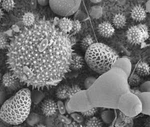
Figure 1. Pollen particles seen with scanning electron microscopy. The outer layers are woody, but contain openings (pores) through which the contents can be digested. (Image courtesy or Louisa Howard, Dartmouth College EM Facility)
Bees get their key nutrients from pollen. There is a lot of nourishment within these woody particles, but they are hard and spiky and their insides are difficult to get at (figure 1).
So the bees’ digestive system has to be able handle this food source. This article looks at the anatomy of the organ where most of this digestion happens, the ventriculus.
Swallowed pollen grains pass along the esophagus to enter the crop. From here they are gathered by the proventricular ‘mouth’ and passed into the ventriculus, through a sleeve, the cardiac (or stomodeal) valve (figure 2) which prevents regurgitation. Within the ventriculus the swallowed pollen is kept within membranes, the peritrophic membranes (figure 3). These thin membranes are secreted by the cells lining the ventriculus (epithelial cells). Nearly all insects have membranes like these, and a considerable amount of study has gone into establishing the role that they play in digestion. In the honey bee these membranes are produced by some of the epithelial cells and successive sheets of membrane peel away from the wall and coat each bolus of pollen as it arrives (figure 3).
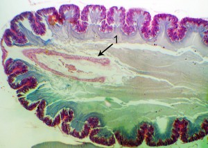 Figure 2. The first part of the ventriculus. The stomodael (or cardiac) valve (1) is a sleeve surrounding the opening bringing food from the crop through the proventriculus. It prevents regurgitation back into the crop. |
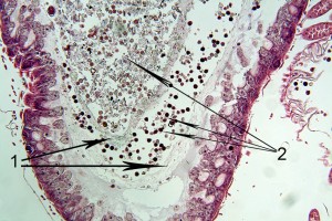 Figure 3. Two peritrophic membranes (1) in the ventriculus. These contain pollen (2) at two different stages of digestion. |
The pollen can move relatively quickly along the length of the ventriculus within the membrane, arriving at the end of the ventriculus within minutes. Here it is generally held for some hours before passing through to the next part of the digestive system which lies beyond the opening of the malphigian tubules, through the pyloric valve, the small intestine (figure 4). Once a layer of peritrophic membrane has peeled away, a further layer is formed, and several layers can be seen in some histological sections through the ventriculus. These peritrophic membranes move more slowly along the ventriculus and are also, ultimately, digested.
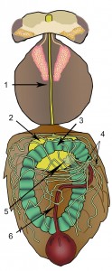
Figure 4. The digestive system. Oesophagus (1), crop (yellow, 2), ventriculus (green, 3), malphigian tubules (4), proventriculus (5), small intestine (red, 6).
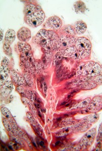
Figure 5. Epithelial cells in the ventriculus laden with packages of digestive enzymes which are seen as small dark particles in the cells.
The membranes were previously thought to provide protection for the lining of the ventriculus from sharp points on the pollen. However, it is more likely that they are important in concentrating a range of digestive chemicals (enzymes) where they are most needed. These enzymes are produced by the epithelial cells themselves and they break down proteins, fats and carbohydrates. The production of enzymes varies along the ventriculus, a higher proportion are released towards the end of the ventriculus (figure 5), whereas in the first part of ventriculus many are bound to the surface of long, finger-like microvilli (figure 6).

Figure 6. Transmission electron microscopy (x5600) image of the surface of a ventricular cell (1). Numerous finger-like villi (2) carrying digestive enzymes extend into the liquid layer over the surface. Further from the surface the liquid is thick with digestive enzymes and particles, the glycocalyx.
The peritrophic membranes probably create compartments within the ventriculus (Moritz and Crailsheim) (figure 7). For example, there is evidence that the enzymes released towards the end of the ventriculus are washed back up the ventriculus in a stream of liquid between the outermost membrane and the ventricular wall. This stream of liquid is likely to come from the malphigian tubules as their flow onwards can be impeded by a ring of muscles at the beginning of the small intestine, the pyloric sphincter. This fluid flowing back up the ventriculus is probably largely absorbed by ventricular cells and returned to the haemolymph from where fluid is once more collected by the malphigian tubules. In this way fluid is recycled between the digestive and excretory systems. In addition a cycle of fluid moves within the ventriculus, forward within the membranes and backwards around the sides (Jimenez and Gilliam). This countercurrent flow system has been observed in other insects.

Figure 7. Diagram of a section through the ventriculus. Pollen grains (colored green) are within compartments made by the various layers of peritrophic membrane (yellow). Fluid containing digestive enzymes flows around the peritrophic membranes back towards the beginning of the
ventriculus.
The enzymes brought back up the ventriculus pass through the membranes and mix with swallowed pollen. In this way enzymes are concentrated within membrane packages where they can be most effective in penetrating pollen and digesting out the contents (figure 8). Once the pollen is finally released from the end of the ventriculus it passes into the small intestine.

Figure 8. Sections through pollen grains from within the small
intestine, after their contents have been digested while passing through the ventriculus.
Polypeptides and other complex molecules released from within the pollen need to be broken down further. The molecules pass through pores in the peritrophic membranes before being cleaved to simpler compounds by enzymes outside the peritrophic membranes and in a viscous layer, the glycocalyx, that coats the epithelial cells, as well as by enzymes bound on the surface of microvilli. Finally, simple compounds are absorbed through the ventricular epithelial cells and released into the haemolymph.
References
Jimenez D.R., Gilliam M. Age-related changes in midgut ultrastructure and trypsin activity in the honey bee, Apis mellifera. Apidologie 1989; 20: 287-303.
Moritz B., Crailsheim K. Physiology of protein digestion in the midgut of the honeybee (Apis mellifera L.). Journal of Insect Physiology 1987 33 923–931