


Anna Gajda DVM, Phd, Warsaw University Of Life Sciences, Institute of Veterinary Medicine, Laboratory of Bee Diseases, anna_gajda@sggw.edu.pl
Ewa Mazur DVM, Warsaw University Of Life Sciences, Institute of Veterinary Medicine, Laboratory of Bee Diseases
Andrzej Bober DVM, National Veterinary Research Institute – State Research Institute, Department of Honeybee Diseases
All life forms, including bees (wild and domesticated species) are threatened by various viruses. They can be singular species-specific or infect a broad range of host (i.e. pollinators). Viral particles are basically a genetic material (RNA or DNA) encapsulated in a protective layer made of protein. Viruses can multiply only in living host cells.
Most viruses are so small that they can be observed only by using highly specialized techniques like electron microscopy (EM). However even in the EM many particles of non-related viruses, that cause very different diseases, look strikingly similiar. That is why the most reliable diagnostic methods are those using serological techniques (i.e. AGID) and molecular biology (i.e. PCR). In some cases the virus can be identified by the symptoms it causes in the colony.
Many viruses, causing severe, often deadly diseases, multiply and spread between single hosts for a long time before causing any visible symptoms. This trait is typical for bee viruses.
The occurence of some viral diseases is dependant on the presence of parasites, i. e. Nosema spp. or Varroa destructor, and others on unfavorable environmental conditions i.e. bad weather making it impossible for bees to fly out, a break in forage sources, etc.).
Many viruses are very commonly present in bee colonies that show no symptoms of infection. There are actually more viruses in bee colonies than any other pathogens. To date 36 bee viruses were found in Apis mellifera colonies. Their phylogenetic origin and target developmental stage of the bee (or the caste) is shown in Table 1.
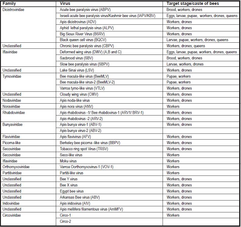
Table 1. Phylogenetic origin of respective viruses and the target stage/caste of bees
Most of those viruses multipliy in bees and brood asymptomatically. Those that pose the biggest threats are described below.
VIRUSES ASSOCIATED WITH VAROOSIS
Deformed wing virus (DWV)
It’s one of the viruses that causes no symptoms in bees and brood without V. destructor. With this parasite (that is a vector and activator of the virus) however DWV can be deadly for individual bees, but often also the entire colony.
By feeding on bees and brood, the mites impair their immunity, which allows the virus to multiply freely. There are emany variants of the virus that differ in virulence (the ability to penetrate, multiply in and damage cells, and in consequence, tissues) in bees. The coexistence of mites and highly virulent viral strains leads to colony deaths. However, regardless of the strain, the longer the mites stay on adult bees, the longer the viral titres, and the more often it leads to emergence of crippled bees from brood cells that the mites entered. Also the more mites in the colony, the more crippled bees emerge.
DWV can be transmitted with royal jelly, sperm (from drones to queen), from queen to egg, from mite to brood and bees.
The symptomatic infection takes many forms from brood death to emergence of crippled bees. Those bees usually have ill developed wings (from gray to brown in color, deformed and shortened), shortened abdomens, movement and orientation impairments. Usually they don’t live past 67 hours. They are removed from the colony earlier and die outside.
It is possible that healthy looking bees will emerge. It is however just apparent health, because those bees have severe nervous system impairments. Their life span is significantly shortened.
Severely infected colonies may rapidly dwindle after feeding for winter and they die shortly after. If the weather allows the bees to fly out, they die in the field, and in the nest only a handful of bees with the queen is left. However, if it is already cold outside, and they cannot fly out, they die in the hive.
DWV is found practically everywhere in the world, but in colonies, in which Varroa control is performed properly (or where Varroa is not yet present), there is no symptoms of this infection.
But in colonies in which V. destructor had a chance to multiply to greater numbers and persist there for extended periods of time, bees with deformed wings begin to appear. The more neglected the colony (treatment wise), the more crippled insects.
Sometimes, after a period of heavy mite infestation, the beekeeper treats the colony well and even kills almost all the parasites. This however does not mean that he gets rid of the virus, because usually DWV manages to multiply so much, that it leads to the colony death anyway, even with a big delay in relation to the treatment.
Acute bee paralysis virus (ABPV)
In the presence of V. destructor this virus can kill both bees and brood. Similar to DWV, the mite impairs the immune response in bees, which allows the virus to multiply freely. V. destructor is also a vector of ABPV.
Heavily infected bees are carriers of the virus and spread it (in huge amounts) with royal jelly and pollen moistened with their saliva to larvae, but also to other adult bees via trophallaxis (food sharing). Transmission of ABPV with food however isn’t usually effective enough for the symptoms to appear. The main role here is played by the mite, that transfers viral partlicles directly into the hemolymph of bees and brood. This route allows fast multiplication of ABPV.
Often the beekeeper doesn’t see any symptoms until the colony dies. It’s because the virus (in the presence of many mites) multiplies very fast, and kills the bees just as rapidly, so the time when the symptoms appear is very short.
Heavily infected bees cannot fly, show symptoms of paralysis and tremors of different magnitude, and in the end they fall off of the combs and are thrown out of the hive by their sister bees.
If it is warm enough so the bees can fly, they usually die in the field leaving food, some brood, a handful of bees and the queen behind.
It may happen that heavily infected bees feed huge ammounts of the virus to larvae with royal jelly, and the brood dies before it is capped. However the beekeeper sees that symptom rather seldom, because the dead larvae are quickly removed by the nurse bees.
Larvae that survive become carriers of ABPV as adult bees and transfer it in big amounts to other larvae during feeding. If, at the same time, there is still a heavy mite infestation, the beekeeper will see foulbrood like symptoms.
Heavily infected colonies usually die in late spring or early Winter.
VIRUSES ASSOCIATED WITH NOSEMOSIS
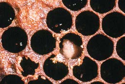
Black queen cell virus (BQCV)
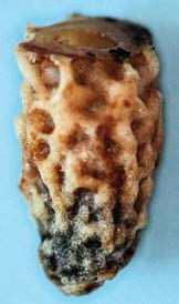
Darkened queen cell. (A. Gajda photo)
This virus plays the key role amongst other viral co-infections with nosemosis. It is transmitted similarly to Nosema spores, via the alimentary route, and is found in considerable ammounts in workers suffering from Nosema infections.
BQCV infection shortens the lifespan of bees significantly. Often they die in early spring, before the new generation of bees can be reared. It results in the course of nosemosis being much more severe and more often leading to colony death (it rapidly dwindles before it can be reinforced with newly emerged bees). Besides the much shorter lifespan, in adult bees there is no other symptoms of BQCV infection.
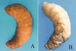
Black Queen Cell Virus – Dead larvae A) yellow, B) with
darkened head. (G. Topolska photo)
Distinctive symptoms can be observed in queen larvae and pupae, however, in natural conditions, when the bees decide about queen rearing on their own, we do not observe this disease. Bees infected with Nosema and BQCV are withdrawn as nurse bees and become foragers faster (they physiologically get older faster).
In contrast, especially in first batches of queen rearing material, there are not enough nurse bees, so infected bees (that normally would be foragers already) still feed the larvae, that is why they can infect queen larvae.
Queen larva or pupa infected with BQCV become pale yellow, it’s cuticle hardens. With time, due to melanin deposition it darkens, leading, by contact, to darkening of the queen cell walls. The virus was named after this specific symptom. One must however remember, that even though dark queen cell walls are commonly associated with BQCV, in reality queen cells with dead queen brood can look completely normal, and contain almost normally looking, pale yellow larvae or pupae inside.
Larvae get infected by eating royal jelly supplied by nurse bees that are carriers of the virus. The larvae get sick only after they have been capped.
During a heavy BQCV infection also worker brood can start dying and the symptoms resemble very closely those of sacbrood disease.
BQCV is a very commonly present virus. Practically in all the apiaries, in which colonies suffer from nosemosis, it can be also found. It is by far the most common cause of queen brood death in queen rearing apiaries.
Bee virus Y (BVY)
It also is transmitted via the alimentary route. It multiplies in the alimentary tract of the bee most efficiently, when the bees are kept in 35oC, whereas when the temperaturę drops even by 5oC, it stops the multiplication completely. BVY shortens the life span of bees and exacerbates the pathogenicity of Nosema spp., however other symptoms were not discovered to date.
Apis mellifera filamentous virus (AmFV)
The particle of this virus is very big compared to most bee viruses, and can be observed in light microscopy (LM), however, recognition can only be done by electron microscopy (EM), because in LM it is visible as a tiny dark speck. AmFV multiplies in fat body and ovaries of infected bees (it is not however transmitted from the queen to the egg).
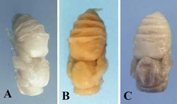
Black Queen Cell Virus Dead pupae A) looking almost normal, B) yellow, C) with darkened front end of the body.
(G. Topolska photo)
Similarly to BQCV and BVY it is transmitted with food. The hemolymph of heavily infected bees becomes milky-white and contains huge ammounts of the virus. In the last phase of the infection, the hemolymph expands its volume and its hemocytes start dissapearing. The influence of AmFV on the course of nosemosis is not as clear as the one of BQCV or BVY. It is commonly thought to be a minimally harmful virus.
Unfortunately, to date there is no available antiviral drug for bees, that is why controlling viral diseases that are associated with other pathogens, consists of this latter pathogen control. It looks similar with viral disease prevention in this case. One should primarily prevent other pathogens from spreading in the hive in order to lower the risk of those viral infections. Namely: proper mite control, hygene (to prevent nosemosis). It is always advised to replace the queen with a young and healthy one, that lays eggs properly, preferably bought from a good breeder, that offers queens producing bees with highly developed hygenic Instinct. It is also known, that endemically occuring bee genotypes always do better with diseases, than the newly introduced ones, that is why one should buy queens from local breeders with long traditions in the region.
VIRUSES NOT ASSOCIATED WITH OTHER PATHOGENS
Chronic bee paralysis virus (CBPV)
It is a very commonly occurring virus.
Just as in case of most viral infections in bees, this one also is often covert, however, with proper conditions occurring, the symptoms start.
CBPV enters hemolymph mostly via wounds from broken hairs on adult bees, but it can also be transmitted via food: with pollen, honey and feces of infected individuals.
The most hairs are broken when during an active beekeeping season the foragers are forced to stay in the hive for too long, which mainly occurs during a sudden brake in forage availability (cold, rainy weather, drought), but also when there are too many colonies in the area (not enough flowers for all the bees). In such conditions CBPV spreads fast in the colony and the symptoms occur in adult bees. However the presence of the virus was confirmed independently of the season in all developmental stages of bees. In very heavily infected colonies also pupae death was observed.
CBPV causes two sets of symptoms (two syndromes). Both lead to death of bees. They can occur simultanously, but always one of them is dominant (it’s genetically conditioned).
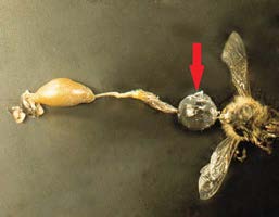
Chronic bee paralysis virus – Alimentary tract and thorax. The arrow shows expanded crop. Spread wings are also depicted. (G. Topolska photo)
Syndrome I. The symptoms result from active multiplication of CBPV in the nervous system of bees, towards which it has the strongest tropism. They are classical progressive paralysis symptoms. Bees tremble unnaturally (body and wings), paralysis of different body parts also occurs, and bees become unable to fly. Sick bees gather in warmer areas of the nest, but later, as useless, they are thrown out of the hive, where they crawl on the ground or up the grass leaves. Their crop is entirely filled with food, which causes the abdomen to be prolonged. Wings are often spread to the sides. The movements of sick bees are wobbly and uncoordinated. Bees die quite fast (but not as fast as in case of ABPV) from the occurence of first symptoms. It often happens in great numbers. The biggest viral loads in this syndrome are found in the crop and salivary glands of sick bees.
Syndrome II – black robbers. Sick workers gradually lose body hair. They become almost black (if the cuticle is black) and shiny, like they were covered in grease. They appear smaller and thinner than healthy bees (body hair makes healthy bees look more chubby). They are attacked by their sister bees and are not let in the hive. Since at the beginning of symptoms they are still able to fly, they circle around the entrance trying to get in. This makes them look like they are trying to rob the colony.
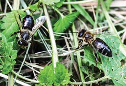
Chronic bee paralysis virus – Black robber bees. (A. Gajda photo)
After a few days symptoms of ataxia and paralysis occur, which later results in death.
The colony usually dies in the middle of Summer. Bees die outside of the hive, in which only a handful of bees with the queen is left. In case of CBPV prevention is based on not letting foragers stay in the hive for too long. It is crucial to provide constant forage, especially in areas where CBPV occurs endemically. For this purpose bees can be transported to forages in other areas or bee friendly crops can be grown near the apiary.
In a sick colony the queen should be replaced with a young one, from a good local breeder. It is also crucial not to place the apiary near forages that already have many hives around.
One absolutely should not try and place crawling/sick looking bees back in the hive! They should immediately be send to the lab to investigate the cause of such symptoms.
Sacbrood virus (SBV)
This virus is pathogenic to brood. Adult bees are only carriers of it. Even though it does not cause symptoms in adults, it can shorten their life. In nurse bees huge amounts of SBV can be found in hypopharyngeal glands. They feed the larvae with their secretion (royal jelly) that contains millions of viral particles. Considerable ammounts of the virus can also be found in pollen stored by the bees. It stays infective for long periods of time in this pollen.
During the first three days of their life, all larvae are fed with royal jelly, that is why they are the most susceptible to SBV infection. If larvae get infected later on, they become carriers of the virus as adults.
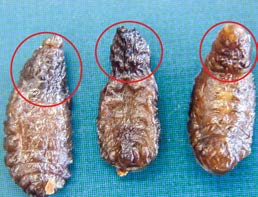
Symptoms however occur only after the larva stretches under the capping. The virus impairs secretion of chitinase, an enzyme responsible for shedding, which prevents the larva from getting rid of old skin, and as a result, from pupating.
Sacbrood Virus – A larva with visibly lifted head.
(G. Topolska photo)
Larva remains in the stretched position with its head lifted. A liquid containing millions of viral particles builds up between the old (unshedded) and the new skin, and the larva resembles a sac with liquid inside, thus the name of the disease.
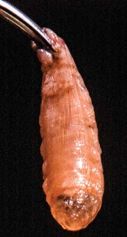
Sacbrood Virus – A larva turned into a sac with liquid.
(G. Topolska photo)
The larva becomes yellow in color, and darkens even more with time (first head becomes brown, then the rest of the body), and dries out taking the shape of a tiny boat (edges and head lifted). Brood becomes scattered on the comb, and the cappings are punctured by the bees that try to remove dead larvae, and get infected in the proces. Hypopharyngeal glands in those bees degenerate, and they physiologically age and become nectar foragers, which prevents them from infecting pollen which is fed to older larvae.
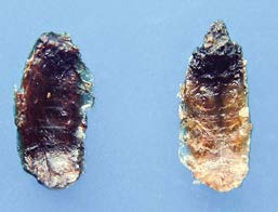
Sacbrood Virus – Dead, dried larvae in the shape of a boat.
(G. Topolska photo)
Sacbrood Virus – Larva with darkened head.
(G. Topolska photo)
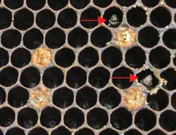
Sacbrood Virus – Scattered brood. Dead larvae with lifted heads are visible as well as a puncture in the cell capping.
(G. Topolska photo)
Symptoms occur in the Spring and usually they retreat spontaneously in Autumn.
Prevention of sacbrood disease consists mostly of ensuring good, constant forage, so the colony can grow evenly and the carrier bees can become foragers. Queens should not be stimulated to lay eggs intensively after forage breaks, because then infected bees that normally would become foragers are forced to feed larvae.
If the disease occurs, the beekeeper should replace the queen and remove the combs on which the brood shows symptoms as well as combs with bee bread (and replace with bee bread from a healthy colony).
DIAGNOSTICS OF VIRAL DISEASES
For many years the diagnosis was only based on the symptoms. It is however now commonly known, that those symptoms may be similar to many diseases and it can lead to misinterpretation, which is dangerous, because most diseases are treated/controlled in very different ways. Hence it is always advised to confirm the preliminary diagnosis with laboratory tests.
There is a spectrum of diagnostic methods to distinguish viral diseases, we are going to describe the ones most widely used.
The AGID test (Agarose Gel Immune Diffusion) is relatively inexpensive and easy to perform, yet not many laboratories perform this test due to difficulties with obtaining proper antisera. It is however recommended as a diagnostic tool here, because it only detects severe infections – the ones which will actually give symptoms and harm the colony.
Rapid development of molecular biology techniques allows also to detect viruses on the genetic level. There are two options: qualitative (end point PCR) – which means it detects the virus without stating the level of infection, and quantitative (real-time PCR) – which tells us how much of the virus is in the sample.
This second method is however usually quite expensive.
A proper sample to detect bee viruses should consist of bees and/or brood that are symptomatic. They should be frozen right after collection and sent to the laboratory with icepacks.